Sindrom Cri Du Chat
 Five P Minus Society
Cri du Chat syndrome

The Cri du Chat syndrome (CdCS) is a genetic disease resulting from a deletion of variable size occurring on the short arm of chromosome 5 (5p-). The incidence ranges from 1:15,000 to 1:50,000 live-born infants. The main clinical features are a high-pitched monochromatic cry, microcephaly, broad nasal bridge, epicanthal folds, micrognathia, abnormal dermatoglyphics, and severe psychomotor and mental retardation. Malformations, although not very frequent, may be present: cardiac, neurological and renal abnormalities, preauricular tags, syndactyly, hypospadias, and cryptorchidism. Molecular cytogenetic analysis has allowed a cytogenetic and phenotypic map of 5p to be defined, even if results from the studies reported up to now are not completely in agreement. Genotype-phenotype correlation studies showed a clinical and cytogenetic variability. The identification of phenotypic subsets associated with a specific size and type of deletion is of diagnostic and prognostic relevance. Specific growth and psychomotor development charts have been established. Two genes, Semaphorin F (SEMAF) and δ-catenin (CTNND2), which have been mapped to the "critical regions", are potentially involved in cerebral development and their deletion may be associated with mental retardation in CdCS patients. Deletion of the telomerase reverse transcriptase (hTERT) gene, localised to 5p15.33, could contribute to the phenotypic changes in CdCS. The critical regions were recently refined by using array comparative genomic hybridisation. The cat-like cry critical region was further narrowed using quantitative polymerase chain reaction (PCR) and three candidate genes were characterised in this region. The diagnosis is based on typical clinical manifestations. Karyotype analysis and, in doubtful cases, FISH analysis will confirm the diagnosis. There is no specific therapy for CdCS but early rehabilitative and educational interventions improve the prognosis and considerable progress has been made in the social adjustment of CdCS patients.
Cri du Chat Syndrome (CdCS) is a genetic disease resulting from a deletion of the short arm of chromosome 5 (5p-). Its clinical and cytogenetic aspects were first described by Lejeune et al. in 1963 [1]. The most important clinical features are a high-pitched cat-like cry (hence the name of the syndrome), distinct facial dysmorphism, microcephaly and severe psychomotor and mental retardation. The size of the deletion ranges from the entire short arm to the region 5p15 [2]. Simmons et al. reported a deletion size ranging from 5 to 40 Mb [3].
CdCS is a rare disease with an incidence ranging from 1:15,000 [4] to 1:50,000 [5] live-born infants. Niebuhr [5] found a prevalence of around 1:350 among over 6,000 mentally retarded people, Duarte et al. [6] found a prevalence of 1:305 among 916 patients attending genetic counselling services and analysed cytogenetically.
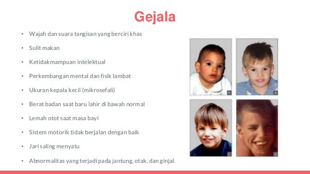
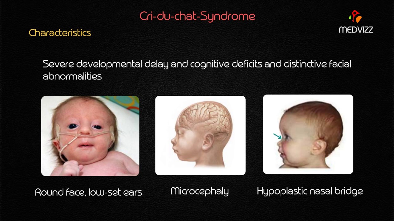
The clinical features at birth are low weight (mean weight 2614 g), microcephaly (mean head circumference 31.8 cm), round face (83.5%), large nasal bridge (87.2%), hypertelorism (81.4%), epicanthal folds (90.2%), downward slanting palpebral fissures (56.9%), down-turned corners of the mouth (81.0%), low-set ears (69.8%), micrognathia (96,7%), abnormal dermatoglyphics (transverse flexion creases) (92%) and the typical cry (95.9%) [1,5,7-19] (percentages from the Italian CdCS Registry [19]) (Fig. 1A,B). Neonatal problems are asphyxia, cyanotic crises, impaired suction and hypotonia. Severe psychomotor retardation becomes evident during the first year of life. Malformations, although not very frequent, may be present: cardiac, neurological and renal abnormalities, preauricular tags, syndactyly, hypospadias, and cryptorchidism. Recurrent respiratory and intestinal infections are reported during the first years of life, although higher sensibility to infections is not reported [20].
The characteristic cat-like cry is probably due to anomalies of the larynx (small, narrow, diamond-shaped) and of the epiglottis (flabby, small, hypotonic), as well as to neurological, structural and functional alterations [5]. Malformations of the cranial base suggest associated anomalies of the brain (rhombencephalic region) and larynx during embryonal development [21].
Specific growth charts for CdCS, based on a multicentre study carried out on 374 patients from the United States, Italy, the United Kingdom and Australia, confirmed the existence of prenatal and postnatal growth retardation [22]. For all ages, median head circumference and weight are near or below the 2nd and 5th percentile, respectively. Height is less affected than weight from birth up to 2 years of age in both sexes. This trend continues until later in life, especially in males. The low weight may be attributed to feeding difficulties and gastroesophageal reflux, both of which are frequent in the first years of life [23]. On the other hand, the slender body shape of many adolescent and adult patients [5,9,14,24] may also be related to the syndrome.
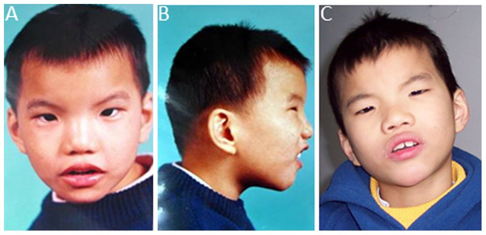
The following features develop with age: the face becomes long and narrow (70.8%), the supra-orbital arch prominent (31.0%), the philtrum short (87.8%), the lower lip full (45.2%), dental malocclusion (open bite) (75.0%) (Fig. 1C,D), palpebral fissures tend to become horizontal (70.2%), divergent strabismus is frequent (44.7%), metacarpi (82.6%) and metatarsi (75.0%) are short resulting in small hands and feet, and prematurely grey hair may be observed (30.4%) [5,7-19,24-28] (percentages from the Italian CdCS Registry [19]).
Myopia and cataract have been reported. Hypersensitivity of the pupil to methacholine and resistance to mydriatics, probably due to a defect of the pupil dilator muscle, have also been described [29,30]. These features have also been found in four patients with Goldenhar's syndrome associated with CdCS [31,32]. Scoliosis, flat foot, pes varus, inguinal hernia and diastasis recti are frequent. Two patients with joint hyperextensibility, skin hyperelasticity and other features of Ehlers-Danlos syndrome [5], and one patient with both clinical manifestations of CdCS and Marfan syndrome have been reported [33]. A patient with a small deletion in 5p15.33 and phenotype suggesting Lujan-Fryns syndrome has been described [34].
Cryptorchidism, sometimes present at birth, is rare in adolescent patients. Sexual development is generally normal in both sexes. A single case of procreation in a CdCS patient (a mother and a daughter with the typical syndrome) has been reported [35].
With age, muscle hypotonia is replaced by hypertonia, and microcephaly becomes more evident. Convulsive crises are rare at all ages. Atrophy of the brainstem mainly involving the pons, cerebellum, median cerebellar peduncles and cerebellar white matter has been revealed by magnetic nuclear resonance imaging [36,37]. A CdCS child with an arachnoid cyst, causing triventricular hydrocephalus by obstruction of the aqueduct of Silvius, has been reported [38]. Metabolic anomalies have been described in CdCS patients: a defect in the synthesis of purine nucleotides (important neuromediators involved in brain development) [39,40] and clinical features associated with non-ketotic hyperglycinaemia, infantile spasms, hypsarrhythmia and brain heterotopia have been reported in a patient with a 5p deletion and typical CdCS [41].
The limited data available about the psychomotor development indicated a severe psychomotor and mental retardation in all patients [5,25]. Prognosis is better for home-reared patients who underwent an early educational program [42-44]. Progress in verbal development is particularly slow [5,45]. Patients' ability to comprehend speech is better than their ability to communicate [46].
A study on psychomotor development was carried out on 91 patients from the Italian Registry [18,47], using the Denver Developmental Screening Test II (DDST II) [48]. This test showed the percentile distribution of patients on the basis of the age of achievement of developmental milestones [47]. A specific psychomotor development chart has been established. Data from the Italian series show that half of the patients walk by themselves at three years old and that all learn to walk later; with regard to the language, 25% of the children are able to make short sentences at 4.5 years old, 50% at 5.5 and almost all the children make short sentences before the age of 10; 50% of the patients feed themselves with a spoon at 3.5 years old and dress at 5 [19]. Although these patients have a range of severe developmental retardation, they can achieve many skills in childhood and continue to learn. This suggests that today's CdCS patients have a better outcome than those in the past [19].
CdCS children have mostly a gentle and affectionate personality. Hyperactivity is present in about 50% of patients and sometimes coexists with aggressiveness, which can be modified with adequate educational programs [5,10,42,49]. The behavioural profile of 27 patients studied by Cornish and Pigram [44] showed self-injury, repetitive movements, hypersensitivity to sounds, clumsiness and obsessive attachment to objects. Hyperactivity and distractibility seems specific to CdCS, if compared to Prader-Willi and Smith-Magenis syndromes [50]. A survey of the prevalence of stereotypy, self-injury and aggression in CdCS children and young adults has been recently carried out by Collins and Cornish [51]. A low level of object-directed behaviour may be an early precursor of hyperactivity, distractibility and stereotypy in older individuals [52]. Nevertheless, early educational interventions and the involvement of families and caregivers allow these behaviours to be improved [19,42].
The introduction of molecular cytogenetic analysis (Fluorescence In Situ Hybridisation, FISH) has allowed the cytogenetic and phenotypic map of 5p to be defined [2,53-56]. Analysis of 80 patients and 148 parents from the Italian Registry of CdCS revealed: a 5p terminal deletion (62 patients: 77.5%), an interstitial deletion (seven patients: 8.75%), a de novo translocation (four patients: 5%), a familial translocation (three patients: 3.75%), a mosaic with two rearranged cell lines (three patients: 3.75%) and a deletion originating from a paternal inversion (one patient: 1.25%). The breakpoints range from p13 to p15.2 (Fig. 2) [56]. This region contains a large number of repetitive sequences that may account for its instability [55,57]. Molecular analysis showed that the deleted chromosome is paternal in most cases: 20/25 (80%) [58], 10/12 (83.3%) [54], 55/61 (90.2%) [56].
The recent studies and observations of Italian patients suggest that partial aneusomy syndromes like CdCS result from abnormal gene dosage (haploinsufficiency) involving a large number of contiguous genes [3,55,56,59]. Other mechanisms, such as gene inactivation due to the position effect or rupture of a very large gene, have also been suggested [60].
A gene for chondrocalcinosis [61] and a gene for asthma [62] have been mapped to 5p15.2. The human Semaphorin F gene (SEMAF) covering at least 10% of this region has been cloned [63]. Due to its role in guiding axons or migrating neuronal precursors during cortical development in mice, it has been suggested that the SEMAF deletion may be responsible for some of the features of CdCS. Another gene, human δ-catenin (CTNND2), has also been mapped to 5p15.2 [59]. δ-catenin is a protein involved in cell motility and is expressed early in neuronal development. δ-catenin deletion seems to correlate with mental retardation in patients with a terminal deletion in this area [59]. δ-catenin knockout mice showed severe impairment of cognitive function, confirming the critical role of this gene in brain function [64].
The results of a recent study in CdCS patients suggest that haploinsufficiency of the telomerase reverse transcriptase (hTERT) gene, localised to 5p15.33, could contribute to the heterogeneous phenotype of CdCS. hTERT is the rate-limiting component for the telomerase activity that is essential for telomere-length maintenance and sustained cell proliferation [65].
Although CdCS is a well-defined clinical entity, individuals with 5p deletion show phenotypic and cytogenetic variability. A few studies, sometimes giving conflicting results, have been performed to correlate the clinical picture with the deletion size [5,24,56,66]. A more severe phenotype and cognitive impairment was reported to be associated with a larger deletion [10,67].
The fact that the phenotype is well recognisable, in spite of the variability in deletion size, has led to the hypothesis that a critical region causes the characteristic clinical picture when present in a hemizygous situation: Niebuhr located this region in a narrow area around 5p15.2 [5,68]. Such an assumption was supported by findings of individuals with a deletion that did not include 5p15.2, who either did not display the typical CdCS phenotype [69,70], or were completely normal [71].
Molecular-cytogenetic analysis allowed Overhauser et al. [2] and Gersh et al. [53] to identify two distinct regions, one for the typical cry in 5p15.3, and another for the other clinical characteristics in 5p15.2. Church et al. [54] distinguished several critical regions: a region for speech retardation, one for the typical cry, one for face dysmorphisms in childhood and one for face dysmorphisms in adulthood (Fig. 2).
A genotype-phenotype correlation study has been carried out in 80 patients from the Italian CdCS Registry. All of them underwent FISH analysis [56]. The results confirmed the importance of deletion of the critical region for manifestation of the CdCS clinical features. However, they also showed a clinical and cytogenetic variability and highlighted a correlation between clinical severity, and the size and type of deletion. In fact, in 62 patients with terminal deletion, the degree of severity (for microcephaly, dysmorphism and psychomotor retardation) has been demonstrated to vary between patients with a small deletion in 5p15.2 and 5p15.1, and patients with a larger deletion. The condition of patients with a deletion in 5p13 appeared particularly severe (Fig. 2).
The variability correlated with the type of deletion in patients with an interstitial deletion, unbalanced translocation resulting in 5p deletion, mosaicism and other rare rearrangements. The study of patients with an interstitial deletion and with a small terminal deletion has enabled the existence of two distinct critical regions (one for dysmorphisms, microcephaly and mental retardation in p15.2, and the other for the typical cry in p15.3) to be confirmed. Moreover, this study allowed the cry region defined by Overhauser et al. [2] to be narrowed distally and supported the hypothesis of a distinct region for speech retardation in p15.3 [54]. Furthermore, two patients who showed an interstitial deletion and a terminal deletion that did not include the critical region and did not show CdCS clinical features, confirmed that not all 5p deletions result in the CdCS phenotype [56,69,70].
In patients with an unbalanced translocation resulting in 5p deletion, the partial trisomy of the other involved chromosome may influence the clinical features, even if the CdCS phenotype prevails [72]. Three patients with mosaicism showed two rearranged cell lines: one with both cell lines deleted, the others with a deleted and a duplicated cell line. In the latter, the CdCS phenotype prevailed over the effect of the partial 5p trisomy present in part of the cells. The patient with the largest duplication had a mild clinical picture, suggesting compensation between deleted and duplicated cell lines [73]. Kitsiou et al. reported a patient with three cell lines in the same tissue: del 5p, dup 5p and a normal one. The mild phenotype in this patient could be mainly due to the normal cell line. However, the duplicated cell line may have contributed to the phenotype through the duplication of the critical CdCS region [73,74].
The deleted chromosome was mainly of paternal origin [54,56,58]: no phenotypic differences caused by imprinting effects were observed in the Italian group of patients [56].
The combination of FISH, comparative genome hybridisation (CGH) and cytogenetic analysis of a patient with dup5q/del5p confirmed that the characteristic cry was due to the deletion at 5p15.3 [75]. Recently Rossi et al. [76], using FISH analysis with bacterial artificial chromosome (BAC) clones in a patient without typical CdCS features, were able to correlate cat-like cry and mild mental retardation with a deletion in 5p15.31, 8.5 Mb away from the short arm telomere. Zhang et al. [77], by using array CGH, refined the CdC critical regions and confirmed the correlation between the severity of mental retardation and the deletion size and type. Using quantitative polymerase chain reaction (PCR), Wu et al. [78] narrowed the critical region for the cat-like cry to a short 640 Kb region and characterised three candidate genes in this region. Harvard et al. [79] found, in a subject with an autism spectrum disorder, a de novo cryptic microdeletion involving 5p15.2.
The identification of phenotypic subsets associated with specific deletions may be of great diagnostic and prognostic relevance. Furthermore, clinical examination combined with molecular analysis of the deletion results in a more personalised evaluation of the patients, which is useful for rehabilitative and educational programs [56].
The diagnosis is first of all clinical, based on typical characteristics such as facial dysmorphisms (facial gestalt), transverse flexion creases, hypotonia in combination with the peculiar cat-like cry. The first test to perform is karyotype analysis, which will confirm the diagnosis. In doubtful cases, when there is a conflict between the clinical suspicion and an apparently normal karyotype result, FISH analysis should be performed [19,34,56,76,80-83].
The importance of FISH for a precise diagnosis of 5p deletions must be emphasised. In the Italian series (80 patients), seven of the patients had not been correctly diagnosed by routine cytogenetics. FISH revealed that five of these patients had an interstitial deletion, one had a small terminal deletion and one had mosaicism [56]. Subtelomeric FISH allows 5p cryptic chromosomal rearrangements to be found [34,82]. Recent techniques, such as array CGH and quantitative PCR, mainly used for research purposes, allow a more precise definition of breakpoints and microrearrangements [77-79].
The clinical features of CdCS patients are not specific if considered separately but, if valued as a whole, they result in a distinct phenotype which, together with the peculiar cry, allows the diagnosis to be suspected at birth. Karyotype analysis of the peripheral blood will confirm the diagnosis.
In the mild cases that can escape the diagnosis or in older patients, it will be the clinical picture (and, above all, the voice that remains abnormal) and the psychomotor retardation that will lead to carrying out of cytogenetic and molecular cytogenetic analyses.
The risk of recurrence is practically negligible for the cases of a de novo deletion, which are the most frequent. However, the possibility of gonadal mosaicism in one of the parents cannot be excluded, even if no recurrence has been reported up to now. It is higher for cases of balanced familial translocation. The reproductive risk for carriers of translocations involving 5p has been defined by evaluation of personal and reviewed data from 54 pedigrees [72]. The same study showed that the risk of unbalanced offspring (according to the pachytene configuration and 5p breakpoint localisation) ranged from 8.7% to 18.8%. The risk for male and female carriers was similar [72]. In these cases, prenatal diagnosis is appropriate.
Prenatal diagnosis by cytogenetic and molecular cytogenetic analyses has been reported in some cases with previous CdCS child, in which the syndrome resulted from a familial balanced translocation [84-88]. Prenatal diagnosis of de novo 5p deletions is not frequent. In two cases it has been performed on the basis of a nonimmune foetal hydrops [89,90], and in another, on the basis of an abnormal ultrasound finding of isolated moderate bilateral ventriculomegaly [91]. Foetal choroid plexus cysts and/or abnormal maternal serum human chorionic gonadotropin (hCG) values in association with CdCS have been reported [92-95]. Chen et al. reported prenatal diagnosis of a foetus with 5p-mosaicism in a case involving advanced maternal age and carried out a review of the literature [88]. In their patient, the mosaic distal 5p deletion was found in association with sonographic markers such as microcephaly and cerebellar hypoplasia [88]. Prenatal diagnosis of the 5p deletion in association with Dandy-Walker syndrome and agenesis of the corpus callosum has been reported [96].
However, it should be noted that not all 5p deletions result in the CdCS phenotype: subjects with short terminal deletions in 5p15.3 may show only a mild or moderate psychomotor retardation [69,70,76,97,98]. Moreover, an interstitial and apparently unbalanced deletion in 5p14, detected by prenatal diagnosis indicated for advanced maternal age and traced through six individuals in three generations, resulted in a completely normal phenotype [71].
There is no specific treatment for CdCS as the cerebral damage resulting from the mutation occurs in the early stages of the embryonal development. Nevertheless, patients benefit from rehabilitative programs, which should be started as soon as possible and involve close collaboration with families, who must be supported psychologically. Moreover, it is important to give to the families updated information about the syndrome, also available through CdCS Support Groups.
Neonatal problems can generally be treated in neonatal pathology departments and intensive treatment is rarely necessary. Breast feeding is possible. For newborns with difficulties in suction and swallowing, physical therapy should start in the first weeks of life. If malformations are present, neonatologists and paediatricians should suggest diagnostic investigations and specialist examinations. It is important to highlight the risk of anaesthesiological problems (intubation difficulties) linked to larynx and epiglottis malformations [99,100]. Intubation difficulties were observed in three cases in the Italian series, but at an older age many patients underwent general anaesthesia without complications [19].
Early rehabilitation (physical therapy, psychomotricity, speech therapy) is recommended for the neurological problems such as psychomotor and speech retardation. As some patients have sensory-neural deafness and speech retardation, audiometric examination should be carried out on all CdCS children. All advised vaccinations are recommended.
Upbringing and rehabilitation are equally important for improvement of the social adaptation of the patients. Guidelines for treatment and follow-up have been reviewed elsewhere [17-19,101].
After the first years of life, the survival expectation is high and morbidity is low. The mortality in the series studied by Niebuhr was about 10%, 75% of which occurred during the first months of life, and up to 90% within the first year [5]. Among the cases described in this study, three patients have lived to be over 50 years of age. Updated data have been reported in a recent study on the natural history of CdCS in a large series of Italian patients [19]. Recent improvements in management of patients with CdCS, with the application of rehabilitative programs, have led to increased psychomotor development, improved autonomy and better social adaptation [19].
The author wishes to thank Telethon Italia (project E.511), "A.B.C. Associazione Bambini Cri du Chat" and Fondazione Cassa di Risparmio di Vercelli for their support, and the research assistants Chiara Castronovo, Stefania Tamiazzo, Michela Godi and Elena Favaron for their collaboration. Moreover the author heartily thanks Mrs Renata Mayer for her generous support in memory of her son Luigi.
The following colleagues collaborated in providing patients, material and clinical information: G. Andria (Napoli), A. Baraldi (Brescia), L. Boggi (Massa Carrara), C. Borrone (Genova), C. Brambilla (Milano), M. Cammarata (Palermo), D. Caufin (Pordenone), M.L. Cavaliere (Napoli), L. Chessa (Roma), F. Dagna Bricarelli (Genova), E. D'Alessandro (L'Aquila), B. Dallapiccola (Roma), A. Di Comite (Taranto), M. Farina (Lamezia Terme), P. Franceschini (Torino), A. Fresia (Vercelli), A. Garau (Cagliari), L. Garavelli (Reggio Emilia), G. Gemme (Genova), A. Giannotti (Roma), M. L. Giovannucci (Firenze), L. Giuffrè (Palermo), A. Guala (Borgosesia), R. Lingeri (Como), A. Lomangino (Bari), A. Lumini (Pistoia), R. Magistrelli (Ancona), M. Martinazzi (Gallarate), T. Mattina (Catania), F. Mollica (Catania), G. Pagano (Como), M. Pagano (Roma), G. Palka (Chieti), G. Pastore (Novara), M. Pergola (Roma), M. Pierluigi (Genova), M.G. Pirastru (Sassari), G. Presta (Brindisi), S. Provera (Vercelli), M.M. Rinaldi (Napoli), G. Rovetta (Manerbio), B. Sacher (S. Daniele del Friuli), M. Stabile (Napoli), A. Selicorni (Milano), L. Tarani (Roma), E. Tarantino (Pisa), R. Tenconi (Padova), E. Valletta (Verona), V. Ventruto (Napoli), M.G. Vianello (Genova), P. Vignetti (Roma), N. Weber (Trieste), L. Zelante (S. Giovanni Rotondo).
- Lejeune J, Lafourcade J, Berger R, Vialatte J, Boeswillwald M, Seringe P, Turpin R. Trois cas de délétion partielle du bras court d'un chromosome 5. CR Acad Sci (D) 1963;257:3098–3102. [PubMed] [Google Scholar]
- Overhauser J, Huang X, Gersh M, Wilson W, McMahon J, Bengtsson U, Rojas K, Meyer M, Wasmuth JJ. Molecular and phenotypic mapping of the short arm of chromosome 5: sublocalization of the critical region for the cri-du-chat syndrome. Hum Mol Genet. 1994;3:247–252. [PubMed] [Google Scholar]
- Simmons AD, Goodard SA, Gallardo TD, Overhauser J, Lovett M. Five novel genes from the cri-du-chat critical region isolated by direct selection. Hum Mol Genet. 1995;4:295–302. [PubMed] [Google Scholar]
- Higurashi M, Oda M, Iijima K, Iijima S, Takeshita T, Watanabe N, Yoneyama K. Livebirths prevalence and follow-up of malformation syndromes in 27,472 newborns. Brain Dev. 1990;12:770–773. [PubMed] [Google Scholar]
- Niebuhr E. The cri du chat syndrome. Epidemiology, cytogenetics and clinical features. Hum Genet. 1978;44:227–275. doi: 10.1007/BF00394291. [PubMed] [CrossRef] [Google Scholar]
- Duarte AC, Cunha E, Roth JM, Ferriera FL, Garcias GL, Martino-Roth MG. Cytogenetics of genetic counseling patients in Pelotas, Rio Grande do Sul, Brazil. Genet Mol Res. 2004;3:303–308. [PubMed] [Google Scholar]
- Dallapiccola B. La patologia cromosomica – Atti dei Congressi della Società Italiana di Medicina Interna, 74° Congresso, Montecatini, 21–24 ottobre. Roma: L. Pozzi; 1973. Malattia del "cri du chat" (5p-) pp. 416–436. [Google Scholar]
- Dallapiccola B, Pistocchi G, Forabosco A, Capra L. Skeletal changes in the "cri du chat" syndrome. Acta Genet Med Gemellol. 1973;22:39–44. [PubMed] [Google Scholar]
- Cerruti Mainardi P, Vianello MG, Bonioli E. Considerazioni su 5 casi di sindrome di "cri du chat". Minerva Pediatr. 1976;28:2389–2400. [PubMed] [Google Scholar]
- Wilkins LE, Brown JA, Nance WE, Wolf B. Clinical heterogeneity in 80 home-reared children with cri-du-chat syndrome. J Pediatr. 1983;102:528–533. doi: 10.1016/S0022-3476(83)80179-6. [PubMed] [CrossRef] [Google Scholar]
- Schinzel A. Catalogue of unbalanced chromosome aberrations in man. Berlin: Walter de Gruyter; 1984. [Google Scholar]
- Benigno V, Cammarata M, Giuffrè L. La sindrome del "cri du chat": dermatoglifi palmari di interesse diagnostico. Minerva Pediatr. 1985;37:251–253. [PubMed] [Google Scholar]
- Fenger K, Niebuhr E. Discriminant analysis of dermatoglyphic sole and palm patterns in Danish cri du chat probands and normal controls. J Ment Defic Res. 1985;29:281–288. [PubMed] [Google Scholar]
- Cerruti Mainardi P. La sindrome del cri du chat in età adulta. In: Andria G, Dagna Bricarelli F, Del Porto G, De Marchi M, Federico A, editor. Patologia genetica ad esordio tardivo. Bologna: Monduzzi; 1987. pp. 113–128. [Google Scholar]
- Bruni L. La sindrome 5p-(sindrome del "cri du chat") In: Vignetti P, Ferrante E, editor. Malattie da aberrazioni cromosomiche. Torino: Edizioni Minerva Medica Italia; 1988. pp. 89–94. [Google Scholar]
- Dallapiccola B. Sindrome del "cri du chat". In: Mastroiacovo P, Dallapiccola B, Andria G, Camera G, Lungarotti MS, editor. Difetti congeniti e sindromi malformative. Milano: McGraw Hill Libri Italia; 1990. pp. 254–255. [Google Scholar]
- Cerruti Mainardi P, Pastore G, Guala A. Sindrome del cri du chat. In: Balestrazzi P, editor. Linee guida assistenziali nel bambino con sindrome malformativa. Milano: CSH; 1994. pp. 75–90. [Google Scholar]
- Cerruti Mainardi P, Perfumo C, Pastore G, Calì A, Guala A, Biroli E, Liverani ME, Egidi I, Zara F, Zerega G, Overhauser J, Pierluigi M, Dagna Bricarelli F. Cri du Chat Syndrome. Ital J Pediatr. 2001;27:840–850. http://www.ijp.it/articoli/2001/vol6-01/indice6_01.htm. [Google Scholar]
- Cerruti Mainardi P, Pastore G, Castronovo C, Godi M, Guala A, Tamiazzo S, Provera S, Pierluigi M, Dagna Bricarelli F. The natural history of Cri du Chat Syndrome. A report from the Italian Register. Eur J Med Genet. 2006. [PubMed]
- Rizzi M. Tesi di Laurea. Facoltà di Medicina e Chirurgia, Università degli Studi di Milano, Anno Accademico; 1997. Valutazione immunologica in pazienti affetti dalla sindrome del cri du chat 5p- [Google Scholar]
- Kjaer I, Niebuhr E. Studies of cranial base in 23 patients with cri-du-chat syndrome suggest a cranial developmental field involved in the condition. Am J Med Genet. 1999;82:6–14. doi: 10.1002/(SICI)1096-8628(19990101)82:1<6::AID-AJMG2>3.0.CO;2-#. [PubMed] [CrossRef] [Google Scholar]
- Marinescu RC, Cerruti Mainardi P, Collins MR, Kouahou M, Coucourde G, Pastore G, Eaton-Evans J, Overhauser J. Growth charts for cri-du-chat syndrome: an international collaborative study. Am J Med Genet. 2000;94:153–162. doi: 10.1002/1096-8628(20000911)94:2<153::AID-AJMG8>3.0.CO;2-#. [PubMed] [CrossRef] [Google Scholar]
- Collins MS, Eaton-Evans J. Growth study of cri du chat syndrome. Arch Dis Child. 2001;85:337–338. doi: 10.1136/adc.85.4.337. [PMC free article] [PubMed] [CrossRef] [Google Scholar]
- Niebuhr E. Antropometry in the Cri du Chat syndrome. Clin Genet. 1979;16:82–95. [PubMed] [Google Scholar]
- Breg WR, Steele MW, Miller OJ, Warburtonb D, Capoa A, Allerdice PW. The cri du chat syndrome in adolescents and adults: clinical finding in 13 older patients with partial deletion of the short arm of chromosome N° 5 (5p-) J Pediatr. 1970;77:782–791. doi: 10.1016/S0022-3476(70)80236-0. [PubMed] [CrossRef] [Google Scholar]
- Niebuhr E. The cat cry syndrome (5p-) in adolescents and adults. J Ment Defic Res. 1971;15:277–291. [PubMed] [Google Scholar]
- Van Buggenhout GJCM, Pijkels E, Holvoet M, Schaap C, Hamel BCJ, Fryns JP. Cri du Chat Syndrome: changing phenotype in older patients. Am J Med Genet. 2000;90:203–215. doi: 10.1002/(SICI)1096-8628(20000131)90:3<203::AID-AJMG5>3.0.CO;2-A. [PubMed] [CrossRef] [Google Scholar]
- Posmyk R, Panasiuk B, Yatsenko SA, Stankiewicz P, Midro AT. A natural history of a child with monosomy 5p syndrome (Cat-cry/Cri-du-chat syndrome) during the 18 years of follow-up. Genet Couns. 2005;16:17–25. [PubMed] [Google Scholar]
- Howard RO. Ocular abnormalities in the cri du chat syndrome. Am J Ophthalmol. 1972;73:949–954. [PubMed] [Google Scholar]
- Kitsiou-Tzeli S, Dellagrammaticas HD, Papas CB, Ladas ID, Bartsocas CS. Unusual ocular findings in an infant with cri-du-chat syndrome. J Med Genet. 1983;20:304–307. [PMC free article] [PubMed] [Google Scholar]
- Kobrynski L, Chitayat D, Zahed L, McGregor D, Rochon L, Brownstein S, Vekemans M, Albert DL. Trisomy 22 and facioauriculovertebral (Goldenhar) sequence. Am J Med Genet. 1993;46:68–71. doi: 10.1002/ajmg.1320460111. [PubMed] [CrossRef] [Google Scholar]
- Choong YF, Watts P, Little E, Beck L. Goldenhar and cri-du-chat syndromes: a contiguous gene deletion syndrome? J AAPOS. 2003;7:226–227. [PubMed] [Google Scholar]
- McLellan MW, Golden WL, Wilson WG. Marfan and cri du chat syndromes in an 18-month-old child: evidence of phenotype interaction. Clin Genet. 1994;46:319–321. [PubMed] [Google Scholar]
- Stathopulu E, Mackie Ogilvie C, Flinter FA. Terminal deletion of chromosome 5p in a patient with phenotypical features of Lujan-Fryns syndrome. Am J Med Genet A. 2003;119:363–6. doi: 10.1002/ajmg.a.10268. [PubMed] [CrossRef] [Google Scholar]
- Martinez JE, Tuck-Muller CM, Superneau D, Wertelecki W. Fertility and cri du chat syndrome. Clin Genet. 1993;43:212–214. [PubMed] [Google Scholar]
- Tamraz J, Rethoré MO, Lejeune J, Outin C, Goepel R, Stievenart JL, Iba-Zizen MT, Cabanis EA. Morphométrie encéphalique en IRM dans la maladie du cri du chat. A propos de sept patients, avec revue de la littérature. Ann Genet. 1993;36:75–87. [PubMed] [Google Scholar]
- De Michele G, Presta M, Di Salle F, Serra L, Mazzaccara A, Della Rocca G, Ambrosio G, Filla A. Cerebellar vermis hypoplasia in a case of cri-du-chat syndrome. Acta Neurol (Napoli) 1993;15:92–6. [PubMed] [Google Scholar]
- Balci S, Oguz KK. Cri-du-chat syndrome associated with arachnoid cyst causing triventricular hydrocephalus. Clin Dysmorphol. 2001;10:289–290. doi: 10.1097/00019605-200110000-00011. [PubMed] [CrossRef] [Google Scholar]
- Lejeune J, Rethoré MO, Peeters M, de Blois MC, Rabier D, Parvy P, Bardet J, Kamoun P. Maladie du cri du chat: acides aminés plasmatiques et urinaires. Ann Genet. 1990;33:16–20. [PubMed] [Google Scholar]
- Peeters MA, Rethoré MO, Aris L, Megarbane A, Cattaneo F, Lejeune J. Metabolic anomalies in cri du chat syndrome (5p-) lymphocytes and the novo purine synthesis. Ann Genet. 1991;34:219–225. [PubMed] [Google Scholar]
- Tsao CY, Wenger GD, Bartholomew DW. Cri du chat syndrome and complex karyotype in a patient with infantile spasms, hypsarrhythmia, nonketotic hyperglycinemia, and heterotopia. Am J Med Genet A. 2005;134:198–201. doi: 10.1002/ajmg.a.30592. [PubMed] [CrossRef] [Google Scholar]
- Wilkins LE, Brown JA, Wolf B. Psychomotor development in 65 home-reared children with cri-du-chat syndrome. J Pediatr. 1980;97:401–405. doi: 10.1016/S0022-3476(80)80189-2. [PubMed] [CrossRef] [Google Scholar]
- Carlin ME. The improved prognosis in cri-du-chat (5p-) syndrome. In: Fraser W, editor. Procedings of the 8th Congress of International Association for the Scientific Study of Mental Deficiency. Edinburgh: Blackwell; 1990. pp. 64–73. [Google Scholar]
- Cornish KM, Pigram J. Developmental and behavioural characteristics of cri du chat syndrome. Arch Dis Child. 1996;75:448–450. [PMC free article] [PubMed] [Google Scholar]
- Cornish KM, Munir F. Receptive and expressive speech skills in children with cri-du-chat syndrome. J Commun Disord. 1998;31:73–80. doi: 10.1016/S0021-9924(97)00052-X. [PubMed] [CrossRef] [Google Scholar]
- Cornish KM, Bramble D, Munir F, Pigram J. Cognitive functioning in children with typical cri du chat (5p-) syndrome. Dev Med Child Neurol. 1999;41:263–6. doi: 10.1017/S0012162299000559. [PubMed] [CrossRef] [Google Scholar]
- Cerruti Mainardi P, Guala A, Pastore G, Pozzo G, Dagna Bricarelli F, Pierluigi M. Psychomotor development in cri du chat syndrome. Clin Genet. 2000;57:459–461. doi: 10.1034/j.1399-0004.2000.570612.x. [PubMed] [CrossRef] [Google Scholar]
- Frankenburg WK, Dodds JB, Archer P, Shapiro H, Bresnick B. The Denver II: a major revision restandardization of the Denver Developmental Screening Test. Pediatrics. 1992;89:91–97. [PubMed] [Google Scholar]
- Dykens EM, Clarke DJ. Correlates of maladaptive behaviour in individuals with 5p- (cri du chat) syndrome. Dev Med Child Neurol. 1997;39:752–756. [PubMed] [Google Scholar]
- Clarke DJ, Boer H. Problem behaviours associated with deletion Prader-Willi, Smith-Magenis, and Cri du Chat Syndromes. Am J Ment Retard. 1998;103:264–271. doi: 10.1352/0895-8017(1998)103<0264:PBAWDP>2.0.CO;2. [PubMed] [CrossRef] [Google Scholar]
- Collins MS, Cornish K. A survey of the prevalence of stereotypy, self-injury and aggression in children and young adults with Cri du Chat syndrome. J Intellect Disabil Res. 2002;46:133–140. doi: 10.1046/j.1365-2788.2002.00361.x. [PubMed] [CrossRef] [Google Scholar]
- Sarimski K. Early play behaviour in children with 5p- (Cri-du-Chat) syndrome. J Intellect Disabil Res. 2003;47:113–120. doi: 10.1046/j.1365-2788.2003.00448.x. [PubMed] [CrossRef] [Google Scholar]
- Gersh M, Goodart SA, Pasztor LM, Harris DJ, Weiss L, Overhauser J. Evidence for a distinct region causing a cat-like cry in patients with 5p deletions. Am J Hum Genet. 1995;56:1404–1410. [PMC free article] [PubMed] [Google Scholar]
- Church DM, Bengtsson U, Nielsen KV, Wasmuth JJ, Niebuhr E. Molecular definition of deletions of different segments of distal 5p that result in distinct phenotypic features. Am J Hum Genet. 1995;56:1162–1172. [PMC free article] [PubMed] [Google Scholar]
- Church DM, Yang J, Bocian M, Shiang R, Wasmuth JJ. A high-resolution physical and transcript map of the Cri du Chat region of human chromosome 5p. Genome Res. 1997;7:787–801. [PubMed] [Google Scholar]
- Cerruti Mainardi P, Perfumo C, Calì A, Coucourde G, Pastore G, Cavani S, Zara F, Overhauser J, Pierluigi M, Dagna Bricarelli F. Clinical and molecular characterization of 80 patients with 5p deletion: genotype – phenotype correlation. J Med Genet. 2001;38:151–158. doi: 10.1136/jmg.38.3.151. [PMC free article] [PubMed] [CrossRef] [Google Scholar]
- Simmons AD, Overhauser J, Lovett M. Isolation of cDNAs from the Cri-du-chat critical region by direct screening of a chromosome 5-specific cDNA library. Genome Res. 1997;7:118–127. [PubMed] [Google Scholar]
- Overhauser J, McMahon J, Oberlender S, Carlin ME, Niebuhr E, Wasmuth JJ, Lee-chen J. Parental origin of chromosome 5 deletions in the cri du chat syndrome. Am J Med Genet. 1990;37:83–86. doi: 10.1002/ajmg.1320370119. [PubMed] [CrossRef] [Google Scholar]
- Medina M, Marinescu RC, Overhauser J, Kosik SK. Hemizigosity of delta-catenin (CTNND2) is associated with severe mental retardation in cri-du-chat syndrome. Genomics. 2000;63:157–164. doi: 10.1006/geno.1999.6090. [PubMed] [CrossRef] [Google Scholar]
- Overhauser J, Marinescu RC, Cheung M, Simmons A, Wixted D, Robin NH, Lovett M. Mapping of genes within a reduced cri-du-chat critical region [abstract] Am J Hum Genet. 1997;A136:776. [Google Scholar]
- Hughes AE, McGibbon D, Woodward E, Dixey J, Doherty M. Localization of a gene for chondrocalcinosis to chromosome 5p. Hum Mol Genet. 1995;4:1225–1228. [PubMed] [Google Scholar]
- The Collaborative Study of the Genetics of Asthma. A genome-wide search for asthma susceptibility loci in ethnically diverse populations. Nature Genet. 1997;15:389–392. doi: 10.1038/ng0497-389. [PubMed] [CrossRef] [Google Scholar]
- Simmons AD, Pueschel AW, Mc Pherson JD, Overhauser J, Lovett M. Molecular cloning and mapping of human Semaphorin F from the Cri-du-Chat candidate interval. Biochem Biophys Res Com. 1998;242:685–691. doi: 10.1006/bbrc.1997.8027. [PubMed] [CrossRef] [Google Scholar]
- Israely I, Costa RM, Xie CW, Silva AJ, Kosik KS, Liu X. Deletion of the neuron-specific protein delta-catenin leads to severe cognitive and synaptic dysfunction. Curr Biol. 2004;14:1657–1663. doi: 10.1016/j.cub.2004.08.065. [PubMed] [CrossRef] [Google Scholar]
- Zhang A, Zheng C, Hou M, Lindvall C, Li K, Erlandsson F, Björkholm M, Gruber A, Blennow E, Xu D. Deletion of the telomerase reverse transcriptase gene and haploinsufficiency of telomere maintenance in Cri du chat syndrome. Am J Hum Genet. 2003;72:940–948. doi: 10.1086/374565. [PMC free article] [PubMed] [CrossRef] [Google Scholar]
- Marinescu RC, Johnson EI, Dykens EM, Hodapp RM, Overhauser J. No relationship between the size of the deletion and the level of developmental delay in cri-du-chat syndrome. Am J Med Genet. 1999;86:66–70. doi: 10.1002/(SICI)1096-8628(19990903)86:1<71::AID-AJMG14>3.0.CO;2-Y. [PubMed] [CrossRef] [Google Scholar]
- Cornish KM, Cross G, Green A, Willatt L, Bradsh JM. A neuropsychological-genetic profile of atypical cri du chat syndrome: implications for prognosis. J Med Genet. 1999;36:567–570. [PMC free article] [PubMed] [Google Scholar]
- Niebuhr E. Cytologic observations in 35 individuals with a 5p-karyotype. Hum Genet. 1978;42:143–146. doi: 10.1007/BF00283634. [PubMed] [CrossRef] [Google Scholar]
- Baccichetti C. Del(5p) without "cri du chat" phenotype [abstract] Hum Genet. 1982;60:389. doi: 10.1007/BF00569228. [CrossRef] [Google Scholar]
- Baccichetti C, Lenzini E, Artifoni L, Caufin D, Marangoni P. Terminal deletion of the short arm of chromosome 5. Clin Genet. 1988;34:219–223. [PubMed] [Google Scholar]
- Overhauser J, Golbus MS, Schonberg SA, Wasmuth JJ. Molecular analysis of an unbalanced deletion of the short arm of chromosome 5 that produces no phenotype. Am J Hum Genet. 1986;39:1–10. [PMC free article] [PubMed] [Google Scholar]
- Cerruti Mainardi P, Calì A, Guala A, Perfumo C, Liverani ME, Pastore G, Overhauser J, Zara F, Pierluigi M, Dagna Bricarelli F. Phenotype-genotype correlation in 7 patients with 5p/autosome translocations. Risk for carriers of translocations involving 5p [abstract] Am J Hum Genet. 2000;A753:145. [Google Scholar]
- Perfumo C, Cerruti Mainardi P, Calì A, Coucourde G, Zara F, Cavani S, Overhauser J, Dagna Bricarelli F, Pierluigi M. The first three mosaic cri du chat syndrome patients with two rearranged cell lines. J Med Genet. 2000;37:967–972. doi: 10.1136/jmg.37.12.967. [PMC free article] [PubMed] [CrossRef] [Google Scholar]
- Kitsiou S, Kolialexi A, Mavrou A. Mosaic Cri du Chat syndrome in a patient exhibiting three 5p cell lines. Prenat Diagn. 2004;24:578–579. doi: 10.1002/pd.906. [PubMed] [CrossRef] [Google Scholar]
- Levy B, Dunn TM, Kern JH, Hirschhorn K, Kardon NB. Delineation of the dup5q phenotype by molecular cytogenetic analysis in a patient with dup5q/del 5p (cri du chat) Am J Med Genet. 2002;108:192–197. doi: 10.1002/ajmg.10261. [PubMed] [CrossRef] [Google Scholar]
- Rossi E, De Gregori M, Patricelli MG, Pramparo T, Argentiero L, Giglio S, Sosta K, Foresti G, Zuffardi O. 8.5 Mb deletion at distal 5p in a male ascertained for azoospermia. Am J Med Genet A. 2005;133:189–192. doi: 10.1002/ajmg.a.30519. [PubMed] [CrossRef] [Google Scholar]
- Zhang X, Snijders A, Segraves R, Zhang X, Niebuhr A, Albertson D, Yang H, Gray J, Niebuhr E, Bolund L, Pinkel D. High-resolution mapping of genotype-phenotype relationships in cri du chat syndrome using array comparative genomic hybridization. Am J Hum Genet. 2005;76:312–326. doi: 10.1086/427762. [PMC free article] [PubMed] [CrossRef] [Google Scholar]
- Wu Q, Niebuhr E, Yang H, Hansen L. Determination of the 'critical region' for cat-like cry of Cri-du-chat syndrome and analysis of candidate genes by quantitative PCR. Eur J Hum Genet. 2005;13:475–485. doi: 10.1038/sj.ejhg.5201345. [PubMed] [CrossRef] [Google Scholar]
- Harvard C, Malenfant P, Koochek M, Creighton S, Mickelson EC, Holden JJ, Lewis ME, Rajcan-Separovic E. A variant Cri du Chat phenotype and autism spectrum disorder in a subject with de novo cryptic microdeletions involving 5p15.2 and 3p24.3-25 detected using whole genomic array CGH. Clin Genet. 2005;67:341–351. doi: 10.1111/j.1399-0004.2005.00406.x. [PubMed] [CrossRef] [Google Scholar]
- Marinescu RC, Johnson EI, Grady D, Chen XN, Overhauser J. FISH analysis of terminal deletions in patients diagnosed with cri-du-chat syndrome. Clin Genet. 1999;56:282–288. doi: 10.1034/j.1399-0004.1999.560405.x. [PubMed] [CrossRef] [Google Scholar]
- Granzow M, Popp S, Keller M, Holtgreve-Grez H, Brough M, Schoell B, Rauterberg-Ruland I, Hager HD, Tariverdian G, Jauch A. Multiplex FISH telomere integrity assay identifies an unbalanced cryptic translocation der(5)t(3;5)(q27;p15.3) in a family with three mentally retarded individuals. Hum Genet. 2000;107:51–57. doi: 10.1007/s004390050010. [PubMed] [CrossRef] [Google Scholar]
- Ensenauer R, Jalal S, Meyer R, Babovic-Vuksanovic D. Unbalanced cryptic 5p deletion/17p duplication identified by subtelomeric FISH in a family with a boy with chimerism and a balanced t(4;5) Am J Med Genet A. 2004;125:86–91. doi: 10.1002/ajmg.a.20420. [PubMed] [CrossRef] [Google Scholar]
- Kondoh T, Shimokawa O, Harada N, Doi T, Yun C, Gohda Y, Kinoshita F, Matsumoto H, Moriuchi H. Genotype-phenotype correlation of 5p-syndrome: pitfall of diagnosis. J Hum Genet. 2005;50:26–29. doi: 10.1007/s10038-004-0213-9. [PubMed] [CrossRef] [Google Scholar]
- Benn PA, Hsu LYF, Verma RS, Aloiso ML, Reich E, Wishnick M. Prenatal diagnosis of minute 5p-deletion: a cytogenetic problem in detection. Obstet Gynecol. 1987;70:449–452. [PubMed] [Google Scholar]
- Smart RD, Retief AE, Overhauser J. Confirmation of a balanced chromosomal translocation using molecular techniques. Prenat Diagn. 1989;9:505–513. [PubMed] [Google Scholar]
- Bernstein R, Bocain ME, Cain MJ, Bengtsson U, Wasmuth JJ. Identification of a cryptic t(5;7) reciprocal translocation by fluorescent in situ hybridization. Am J Med Genet. 1993;46:77–82. doi: 10.1002/ajmg.1320460113. [PubMed] [CrossRef] [Google Scholar]
- Pettenati MJ, Hayworth R, Cox K, Rao PN. Prenatal detection of cri du chat syndrome on uncultered amniocytes using fluorescence in situ hybridization (FISH) Clin Genet. 1994;45:17–20. [PubMed] [Google Scholar]
- Chen CC, Lee CC, Chang TY, Town DD, Wang W. Prenatal diagnosis of mosaic distal 5p deletion and review of the literature. Prenat Diagn. 2004;24:50–57. doi: 10.1002/pd.794. [PubMed] [CrossRef] [Google Scholar]
- Tullu MS, Muranjan MN, Sharma SV, Sahu DR, Swami SR, Deshmukh CT, Bharucha BA. Cri-du-chat syndrome: clinical profile and prenatal diagnosis. J Postgrad Med. 1998;44:101–104. [PubMed] [Google Scholar]
- Aoky S, Hata T, Hata K, Miyazaki K. Antenatal sonographic features of cri du chat syndrome. Ultrasound Obstet Gynecol. 1999;13:216–219. doi: 10.1046/j.1469-0705.1999.13030216.x. [PubMed] [CrossRef] [Google Scholar]
- Stefanou EG, Hanna G, Foakes A, Crocker M, Fitchett M. Prenatal diagnosis of cri du chat (5p) syndrome in association with isolated moderate bilateral ventriculomegaly. Prenat Diagn. 2002;22:64–66. doi: 10.1002/pd.243. [PubMed] [CrossRef] [Google Scholar]
- Sarno AP, Jr, Polzin WJ, Kalish VB. Fetal choroid plexus in association with cri du chat (5p-) syndrome. Am J Obstet Gynecol. 1993;169:1614–5. [PubMed] [Google Scholar]
- Muller F, Aegerter P, Boue A. Prospective maternal serum chorionic gonadotropin screening for the risk of fetal chromosome anomalies and of subsequent fetal and neonatal deaths. Prenat Diagn. 1993;13:29–43. [PubMed] [Google Scholar]
- Fankhauser L, Brundler AM, Dahoun S. Cri-du-chat syndrome diagnosed by amniocentesis performed due to abnormal maternal serum test. Prenat Diagn. 1998;18:1099–100. doi: 10.1002/(SICI)1097-0223(1998100)18:10<1099::AID-PD400>3.0.CO;2-H. [PubMed] [CrossRef] [Google Scholar]
- Weiss A, Shalev S, Weiner E, Shneor Y, Shalev E. Prenatal diagnosis of 5p deletion syndrome following abnormally low maternal serum human chorionic gonadotrophin. Prenat Diagn. 2003;23:572–574. doi: 10.1002/pd.645. [PubMed] [CrossRef] [Google Scholar]
- Vialard F, Robyr R, Hillion Y, Molina Gomes D, Selva J, Ville Y. Dandy-Walker syndrome and corpus callosum agenesis in 5p deletion. Prenat Diagn. 2005;25:311–313. doi: 10.1002/pd.1130. [PubMed] [CrossRef] [Google Scholar]
- Kushnick T, Rao KW, Lamb AN. Familial 5p-syndrome. Clin Genet. 1984;26:472–476. [PubMed] [Google Scholar]
- Bengtsson U, McMahon J, Quarrel Q, Rubenstein C, David K, Greenberg F, Wasmuth JJ. Phenotypically normal carriers of unbalanced terminal deletion of 5p transmit deletions to offspring who display growth and developmental delay [abstract] Am J Hum Genet. 1990;A47:208. [Google Scholar]
- Yamashita M, Tanioka F, Taniguchi K, Maisuki A, Oyama T. Anesthetic considerations in cri du chat syndrome: a report of three cases. Anesthesiology. 1985;63:201–202. [PubMed] [Google Scholar]
- Brislin RP, Stayer SA, Schwartz RE. Anaesthetic considerations for the patient with cri du chat syndrome. Paediatr Anaesth. 1995;5:139–141. [PubMed] [Google Scholar]
- Cerruti Mainardi P, Medolago LM, Pedrinazzi M. La Sindrome del Cri du Chat. Firenze: Grafiche Borri, S. Casciano V.P; 2002. http://www.criduchat.it/cdc/doc_vari/ABCLibrettoSCDC72/ABClibrettoSCDC72.pdf. [Google Scholar]
Gallery Sindrom Cri Du Chat
 The Cri Du Chat Syndrome Semantic Scholar
 Cri Du Chat Syndrome Bree S Story Penfield Building Blocks
 Apa Itu Sindrom Cri Du Chat Kenali Lebih Dini Pada Balita
 Apa Itu Mutasi Ppt Download
:max_bytes(150000):strip_icc()/ChildWithDiGeorgeSyndrome-5c4f3794c9e77c0001d76209.jpg) An Overview Of Cri Du Chat Syndrome
An Overview Of Cri Du Chat Syndrome
 Cri Du Chat Syndrome
 Essay On Cri Du Chat Syndrome Cri Du Chat Essay Words
 Dubowitz Syndrome Is A Complex Comprised Of Multiple
Baclofen As Related To Cri Du Chat Syndrome Pictures
 Sindrom Macijeg Placa Bionet Skola
 The Cry Of The Cat Cri Du Chat Syndrome Page 6997818
 Angelman Syndrome A Review Of The Clinical And Genetic
 Ciri Penderita Sindrom Cri Du Chat
 Cri Du Chat Sindrom Langka Yang Membuat Suara Anak Seperti
 Diagnosis Of Smith Magenis Syndrome
 Apakah Sindrom Cri Du Chat Penyakit Keturunan
 Forgotten Diseases Research Foundation Cri Du Chat
 Cri Du Chat Cat S Cry Syndrome Symptoms Treatment More
 Sindrom Du Chat Cat S Cry Gejala Rawatan Lagi 2019
Mukwonago Child With Cri Du Chat Syndrome Thrives And Shines


0 Response to "Sindrom Cri Du Chat"
Post a Comment